
Bone Infection Foot Treatment in Porbandar: Curing It Properly
Published: 10 August 2026 · Last Updated: August 2026
Key Takeaways
- Bone infection in the foot, called osteomyelitis, usually spreads inward from a long standing skin ulcer.
- A wound you can touch bone through carries a high probability of bone involvement and needs imaging quickly.
- Plain X rays look normal for the first two to three weeks, so a clear film never rules infection out.
- Deep bone samples identify the true organism, whereas surface swabs mostly grow harmless skin bacteria.
- Dead infected bone has no blood supply, therefore antibiotics cannot reach it and surgery becomes necessary.
- Most feet with restorable circulation are saved when clearance happens before infection spreads widely.
A foot wound that keeps discharging month after month is usually telling you something specific. Bone Infection Foot Treatment in Porbandar is built around that message, because a small opening that refuses to dry up very often sits directly above infected bone. Skin infections settle with antibiotics, whereas infected bone rarely does, and recognising that difference early changes the entire outcome for the foot.
Patients travelling to Dr. Ashutosh Shah often bring a thick folder of prescriptions. Four or five antibiotic courses have been finished, the wound closes briefly and then reopens, and nobody has yet ordered an MRI or taken a bone sample. Frustration by that point runs deep. Once the infected bone is identified and cleared surgically, the same wound frequently heals within weeks.
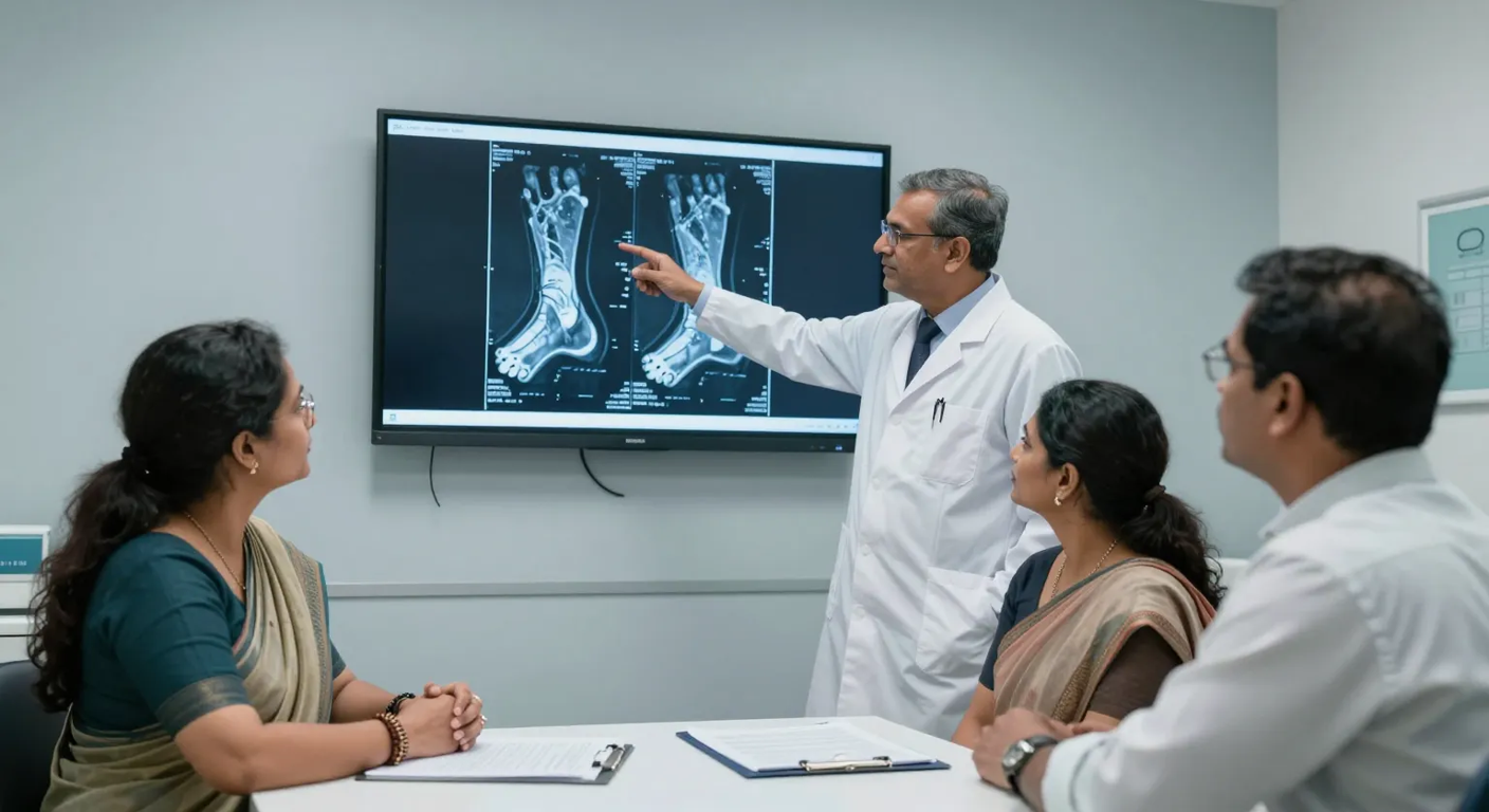
What is bone infection in the foot?
Bacteria that reach bone multiply inside it, pressure builds within the rigid structure, and the tiny vessels feeding segments of bone become blocked. Those segments then die. Surgeons call such a fragment a sequestrum, and it behaves like an infected splinter locked inside the foot.
Osteomyelitis of the foot is bacterial infection within bone and marrow that destroys the local blood supply, leaving dead infected fragments which antibiotics cannot reach and which usually have to be removed surgically.
Toes, the heel and the bones under the ball of the foot are affected most often, since those areas carry pressure and sit directly beneath ulcers. Diabetes, kidney disease, smoking and long term steroid use all raise the risk considerably.
How is bone infection different from a skin infection?
Skin and soft tissue infection produces spreading redness, warmth, swelling and often fever, and it settles within a week or two on suitable antibiotics. Bone infection behaves quietly instead, with a small persistent opening, scanty discharge, minimal fever and a wound that reopens after every treatment course.
Duration separates the two more reliably than appearance. Any foot wound still discharging after six weeks, especially one that has closed and reopened repeatedly, deserves investigation for bone involvement rather than another prescription.
Which warning signs should prompt action?
- A wound through which bone can be felt when probed gently with a sterile instrument.
- A single toe that is swollen, red and sausage shaped along its whole length.
- A small sinus opening that keeps draining thin fluid for weeks despite dressings.
- Repeated infection returning to exactly the same spot after each antibiotic course.
- An ulcer showing no measurable shrinkage after four weeks of otherwise correct wound care.
Diabetic patients rarely feel pain in any of these situations, which is precisely the danger. Neuropathy removes the alarm, so the decision to seek assessment must come from what the foot looks like rather than from how much it hurts.
Which tests confirm bone infection?
Diagnosis combines examination, imaging and laboratory work. Probing the wound to bone raises suspicion strongly, MRI then shows marrow involvement and maps how far infection extends, and blood tests track infection markers alongside sugar control and haemoglobin.
Circulation testing runs in parallel, because a foot with blocked arteries needs blood flow restored before any surgery is attempted. Most importantly, bone taken during surgery is cultured. Surface swabs identify organisms sitting on the wound rather than the bacteria living inside the bone, so treatment guided by a swab frequently targets the wrong thing entirely.
Why do antibiotics alone keep failing?
Antibiotics travel through blood. Dead bone has no blood supply whatsoever, therefore no medicine reaches the bacteria hiding inside it, regardless of dose or duration. Each course suppresses surrounding infection temporarily, the wound looks better briefly, then bacteria emerge again from the protected fragment.
Removing that fragment changes the arithmetic completely. Once dead bone is gone and healthy bleeding bone remains, antibiotics finally reach every remaining organism, and a defined course then finishes the job. Research collected on PubMed consistently reports higher cure rates when surgical clearance is combined with culture guided antibiotics rather than medicines alone.
Treatment options explained
- Surgical clearance: infected and dead bone is removed until healthy bleeding bone is reached, forming the foundation of any real cure.
- Culture guided antibiotics: a defined course, commonly four to six weeks, follows surgery and is narrowed once bone culture results arrive.
- Local antibiotic carriers: beads or cement placed into the cavity deliver high concentration medicine exactly where blood flow is limited.
- Restoring circulation: angioplasty or bypass precedes bone surgery whenever Doppler shows inadequate arterial supply.
- Soft tissue cover: grafts or flaps close the defect and bring fresh blood supply across the cleared area.
- Limited amputation: removing one infected toe or ray sometimes saves the whole foot and preserves normal walking.
Grafting and flap results after bone clearance can be seen in the before and after gallery.
How Bone Infection Foot Treatment in Porbandar is sequenced
- Detailed assessment: wound duration, previous antibiotics, diabetes control and circulation are reviewed, then the ulcer is probed for bone contact.
- Imaging and blood work: MRI or X ray, infection markers, sugar profile and Doppler studies are completed together to avoid repeated journeys.
- Circulation first: arterial blockage is treated before bone surgery whenever flow proves inadequate, since clearance without blood supply simply fails.
- Bone clearance with culture: infected bone is removed to healthy margins while deep samples go for culture and sensitivity.
- Targeted antibiotic course: medicines are narrowed once results arrive, then continued for the planned duration with monitoring.
- Closure and review: the defect is grafted or flap covered, after which scheduled reviews confirm that markers and the wound both settle.
Travelling to Surat from Porbandar
Elegance Clinic runs only at Adajan and Vesu in Surat, and no branch, camp or visiting centre exists in Porbandar. Saying so plainly helps families plan properly. Porbandar sits roughly eight to nine hours away by road, therefore a single well organised visit with all reports gathered beforehand is far kinder than repeated trips.
Local hospitals manage acute infections, drainage and routine dressings capably, so travel is worth weighing in defined situations: a wound open beyond six weeks, bone visible or palpable in the ulcer, infection returning repeatedly to the same site, or an amputation recommendation the family wishes reviewed. The outstation patients guide covers appointment scheduling, reports and accommodation, while MRI films and culture reports shared through the clinic contact page allow most planning to finish before you set out. Follow up reviews then continue by video, with dressings done locally.
What does treatment cost?
Cost follows the extent of surgery and the length of antibiotic treatment. A single infected toe cleared as a day procedure sits at the lower end, whereas wide clearance combined with angioplasty, intravenous antibiotics and flap reconstruction costs considerably more. Hospital stay and diabetes management influence the total as well.
A written estimate follows assessment, listing surgery, imaging, medicines, dressings and review visits. Because treatment commonly continues across six to eight weeks, instalment options help families budget without pausing care, and the EMI and financing guide sets out how those plans work.
Myths that waste months
- Stronger or longer antibiotic courses cannot cure established bone infection, since medicine never reaches dead bone.
- A normal X ray does not exclude the diagnosis, because bone changes appear only weeks after infection starts.
- Painless wounds are not minor wounds, given that diabetic neuropathy removes the warning entirely.
- Surgery is not a last resort here, as early clearance preserves far more foot than delayed clearance does.
- Home remedies and repeated dressing changes cannot reach infection sitting inside bone.
Aftercare and preventing recurrence
Recurrence risk persists for years, so aftercare deserves genuine commitment. Finish the entire antibiotic course exactly as prescribed, hold blood sugar within target daily, and attend every scheduled review even when the foot looks perfectly healed. Offloading footwear or custom insoles then keep pressure away from the treated area.
Daily inspection catches trouble while it remains trivial. Check the sole and between the toes each evening, using a mirror where sensation is reduced. Should fresh discharge, swelling or a new opening appear, early review through online consultation booking prevents another long treatment cycle.
Why patients choose Elegance Clinic
Families select Elegance Clinic because bone clearance, wound management and reconstruction are handled by one plastic surgeon within a single plan rather than passed between departments. Dr. Ashutosh Shah brings more than twenty two years of reconstructive experience across debridement, skin grafting, flap cover and diabetic foot salvage, which keeps the sequence moving without avoidable gaps.
Anyone arranging Bone Infection Foot Treatment in Porbandar should put two questions to any treating team. Ask whether a bone sample has been cultured rather than a surface swab, and ask whether circulation has been formally tested before surgery is planned. Those two answers separate treatment that cures from treatment that merely postpones.
FAQs
Can bone infection be cured without surgery?
Very early infection caught within days occasionally settles on antibiotics alone. Established infection containing dead bone rarely does, because no blood supply carries medicine there. Surgical removal followed by a targeted course offers a far higher cure rate.
How long does the antibiotic course last?
Four to six weeks is typical after clearance, frequently starting with intravenous medicines before switching to tablets. Exact duration depends on the organism identified, how much bone was removed and how infection markers respond during monitoring.
Does a clear X ray mean the bone is healthy?
No. Visible changes take roughly two to three weeks of infection to appear, so early disease looks entirely normal on film. MRI detects marrow involvement much sooner and shows precisely how far infection extends.
Will the whole foot need to be removed?
Usually not. Major amputation becomes likely only when circulation cannot be restored or when infection threatens life. Timely clearance with adequate blood flow and good sugar control saves the majority of affected feet.
Why does the small opening keep draining?
Persistent drainage from a tiny sinus almost always indicates dead infected bone acting as a reservoir underneath. That tract continues discharging until the fragment is surgically removed, however many antibiotic courses are completed first.
How soon can I walk after the operation?
Protected walking often begins within a few days using special offloading footwear or a boot that keeps pressure off the operated area. Unrestricted walking waits until the wound has healed and the surgeon confirms it is safe.
Is the surgery done under general anaesthesia?
Small procedures on a single toe are frequently performed under regional or local anaesthesia as day cases. Extensive clearance or flap reconstruction generally needs spinal or general anaesthesia with a short hospital stay afterwards.
Can infection return after successful treatment?
Recurrence is possible, particularly where pressure returns to the same spot or sugar control slips. Consistent offloading footwear, daily foot inspection and regular reviews reduce that risk substantially over the following years.
Ready to discuss your concern in person? Book a private consultation at our Adajan or Vesu clinic. Call +91 83205 00350 or use the Book Consultation button.